
Axe 1 : Approches technologiques et méthodologiques multi-échelles pour une santé de précision :
Development of alternative Organ-on-Chip (OoC) models
A new Microfluidic platform to follow the extravasation process and collect extravasated cancer cells
To prevent metastatic recurrences, we need to understand the steps of metastases that occur during and after dissemination of tumor cells from the primary site and develop strategies to block these steps. Once arrested on the capillary beds of targeted organs, tumor cells have to extravasate through the endothelial wall and enter these organs. This extravasation step is crucial for the establishment of metastatic tumors. One point that needs to be elucidated is the stemness characteristics of metastatic cancer cells.
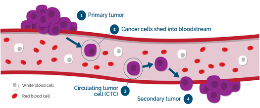 The first hypothesis is that cancer cells need to be in a stem-ness state to be able to initiate extravasation and then generate metastasis. The other hypothesis is that cancer cells will only display stemness characteristics once extravasated in the appropriate microenvironment.
![]()
 Devices and results ________________
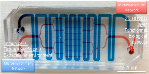
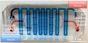
We developed an original biomimetic platform which enable not only to follow in real-time the extravasation process but also to collect, for further analyses, extravasated cancer cells. We developed an original biomimetic microchip with a specific new design allowing to study and follow in real time the cancer cells and their interactions with the endothelium wall.
![]()
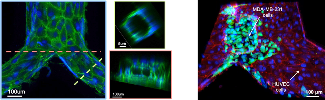 Our device enables to collect extravasated cancer cells through the bottom collecting chamber and compare them, in term of phenotype with those that were not able to extravasate. We use time-lapse imaging in a biomimetic microfluidic device to study the metastatic potential of breast cancer cells focusing on CSCs/non-CSCs. This new original microfluidic microchip enables to study in real-time cancer cells and their interaction with the endothelium through a complex bifurcation network mimicking capillary bed. The effect of the metastatic microenvironment is studied, through the modulation of the bottom collecting chamber. In this study, we developed a new microfluidic device in order to study tumor cell extravasation.
Contact : anthony.treizebre ![]() univ-lille.fr
univ-lille.fr
![]()
Collaborations

Biosensors for studying and quantifying the infectious nature of parasites
Biosensors for studying and quantifying the infectious nature of parasites
Les récentes épidémies de Covid-19 et de grippes aviaires ont mis en lumière les risque de transmission d’agents pathogènes de l’animal à l’Homme (maladie dites « zoonotiques »). Ces crises sanitaires ont également mis en évidence l’importance de définir le plus tôt possible la capacité de différents variants, ou souches, à induire la maladie (notion de la virulence).
Une fois déterminée sa virulence, la gestion des risques liés à un agent infectieux passe par le déploiement de nouvelles stratégies thérapeutiques et la recherche de nouveaux médicaments.
Dans ce contexte, les parasites unicellulaires appartenant au genre Cryptosporidium représentent un défi particulièrement complexe à relever aussi bien pour les instances de santé public que pour les entreprises pharmaceutiques. En effet, malgré des décennies de recherche, il demeure toujours de vraies lacunes technologiques dans les modèles de culture in vitro mis à disposition des laboratoires de recherche. De même l’absence de médicaments permettant de traiter les patients immunodéprimés contaminés par ce parasite limite la capacité des praticiens hospitaliers ou des vétérinaires à contrôler les épidémies de Cryptosporidiose (la maladie induite par ce type de parasites).
Pour répondre à ces grands enjeux de la médecine animale et humaine, des stratégies transdisciplinaires ont été élaborées. Elles combinent les savoirs faire issus de la microélectronique (construction d’outils miniaturisés en salle blanche), de la biologie et de l’intelligence artificielle.
Grace à cette approche transdisciplinaire, une plateforme de criblage de médicaments s’appuyant sur des mesures électriques par spectroscopie d’impédance a été développée. Cet outil a été élaboré dans le cadre d’un projet Européen (porté par l’un membre de l’équipe BioMEMS). Ce dispositif d’analyse est basé sur un réseau de microélectrodes interdigitées sur lesquelles des cellules humaines sont cultivées et infectées par le parasite (C. parvum). Les données obtenues ont montré que la réponse électrique enregistrée était modifiée en présence de molécules inhibant le développement du parasite. Ces résultats représentent la première preuve de concept montrant l’utilisation d’une méthode d’analyse électrique non invasive utilisée à des fins de criblage de molécules à visée thérapeutique dirigée contre un parasite. Cette stratégie ouvre la voie à l’utilisation en routine de la spectroscopie d’impédance dans le criblage de futurs médicaments de façon automatisée et cinétique.
Deux questions sont maintenant en cours d’étude sur cette plateforme impédimétrique :
- Peut elle mesurer des variations de signature impédimétrique en fonction de souches de parasite présentant des pouvoir infectieux différents ?
- Peut elle être appliquée à d’autres agents pathogènes tels que des virus ?
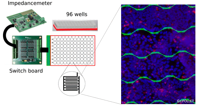
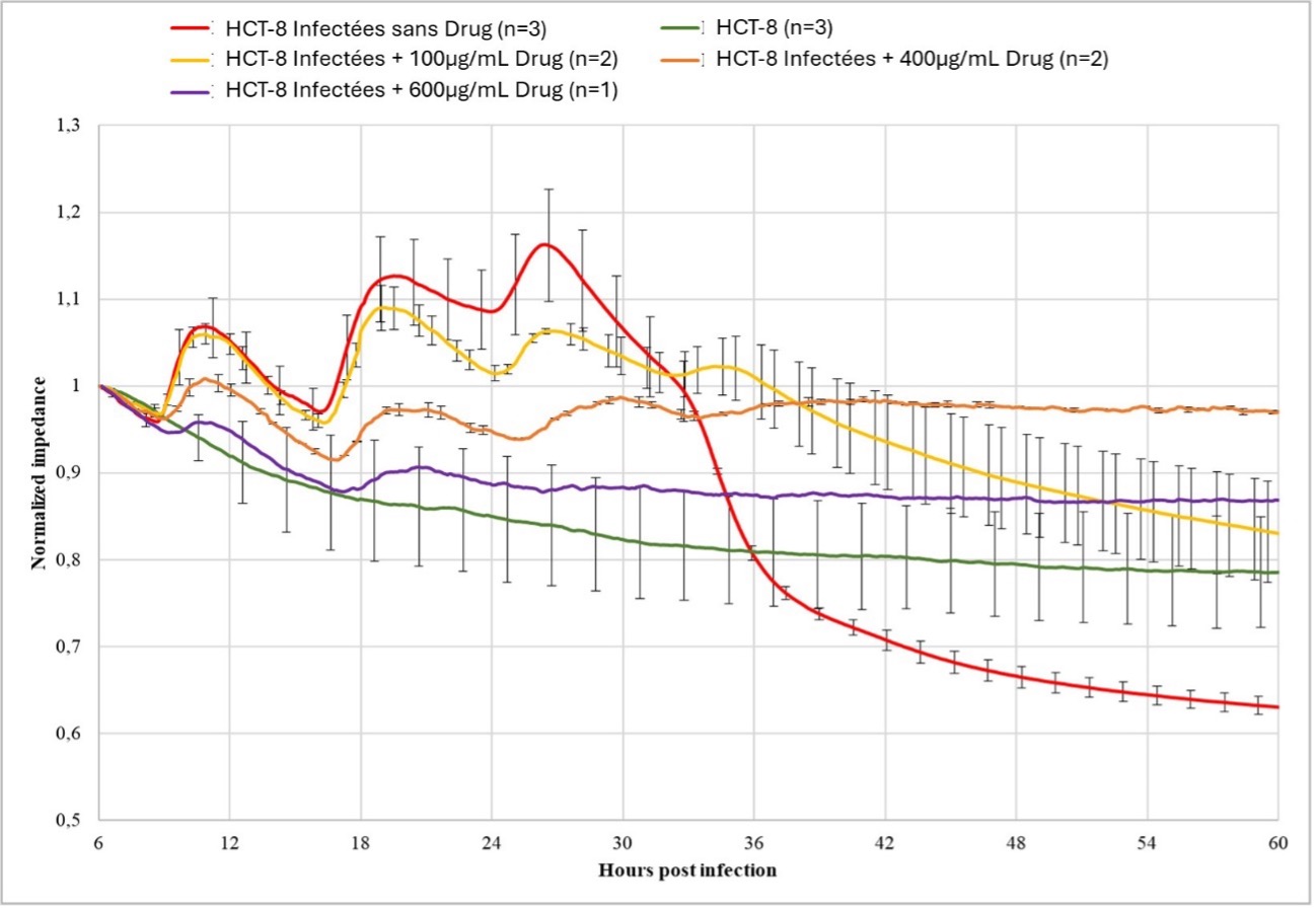
Droite: Conception générale du dispositif de criblage de médicaments. L’image de droite montre des cellules HCT-8 dont le noyau est coloré au DAPI (points bleus) et qui se développent sur des microélectrodes interdigitées. Cette couche cellulaire est infectée par C. parvum (souche IOWA) marqué par des anticorps anti-Cryptosporidium marqués au Cy3 (points rouges). Gauche: Variation de la valeur d’impédances en fonction du temps mesurée sur des cellules Humaines (HCT-8) infectées par des parasites. Les données sont enregistrées varient en fonction d’une gamme de concentration d’un composé présentant une activité inhibitrice. En vert : Témoin négatif comprenant les cellules seules non infectées. En Rouge, témoin positif comprenant des cellules infectées avec des parasites en l’absence de molécule inhibitrices. En Jaune, orange et violet, cellules infectées avec l’ajout une concentration finale de molécules inhibitrice respectivement de 100 µg/mL, 400 µg/mL et 600 µg/mL. Les valeurs présentées correspondent à la moyenne des résultats obtenus sur n expériences +/- la déviation standard.
Contact : jerome.follet ![]() yncrea.fr
yncrea.fr
Contact : alexis.vlandas ![]() univ-lille.fr
univ-lille.fr
Publications ________________
- Lenière AC, Vlandas A, Follet J. Treating cryptosporidiosis: A review on drug discovery strategies. Int J Parasitol Drugs Drug Resist. 2024 Apr 20;25:100542. doi: 10.1016/j.ijpddr.2024.100542. Epub ahead of print. PMID: 38669849; PMCID: PMC11066572.
- Hoque S, Pinto P, Ribeiro CA, Canniere E, Daandels Y, Dellevoet M, Bourgeois A, Hammouma O, Hunter P, Gentekaki E, Kváč M, Follet J, Tsaousis AD. Follow-up investigation into Cryptosporidium prevalence and transmission in Western European dairy farms. Vet Parasitol. 2023 Jun;318:109920. doi: 10.1016/j.vetpar.2023.109920. Epub 2023 Apr 1. PMID: 37030025.
- Pinto P, Ribeiro CA, Hoque S, Hammouma O, Leruste H, Détriché S, Canniere E, Daandels Y, Dellevoet M, Roemen J, Barbier Bourgeois A, Kváč M, Follet J, Tsaousis AD. Cross-Border Investigations on the Prevalence and Transmission Dynamics of Cryptosporidium Species in Dairy Cattle Farms in Western Mainland Europe. Microorganisms. 2021 Nov 20;9(11):2394. doi: 10.3390/microorganisms9112394. PMID: 34835519; PMCID: PMC8617893.
- Baydoun M, Vanneste SB, Creusy C, Guyot K, Gantois N, Chabe M, Delaire B, Mouray A, Baydoun A, Forzy G, Chieux V, Gosset P, Senez V, Viscogliosi E, Follet J, Certad G. Three-dimensional (3D) culture of adult murine colon as an in vitro model of cryptosporidiosis: Proof of concept. Sci Rep. 2017 Dec 11;7(1):17288. doi: 10.1038/s41598-017-17304-2. PMID: 29230047; PMCID: PMC5725449.
- Dibao-Dina A, Follet J, Ibrahim M, Vlandas A, Senez V. Electrical impedance sensor for quantitative monitoring of infection processes on HCT-8 cells by the waterborne parasite Cryptosporidium. Biosens Bioelectron. 2015 Apr 15;66:69-76. doi: 10.1016/j.bios.2014.11.009. Epub 2014 Nov 10. PMID: 25460884.
On going projects ___________
 Projet Viamea, financé par le programme « Stimule », région Hauts de France
Projet Cryptostrain, financé par demi bourse de thèse région Hauts de France et Junia
Projet Healthy Teeth, finance par le programme Interreg France Wallonie Flandres (FWVl)

Collaborations
Innovative microsystems for the multi-physical characterisation of living cells
Distinguishing cancer cells based on their biophysical signatures
The BioMEMS team has been developing advance bioMEMS devices for cancer cell analysis in two axes:
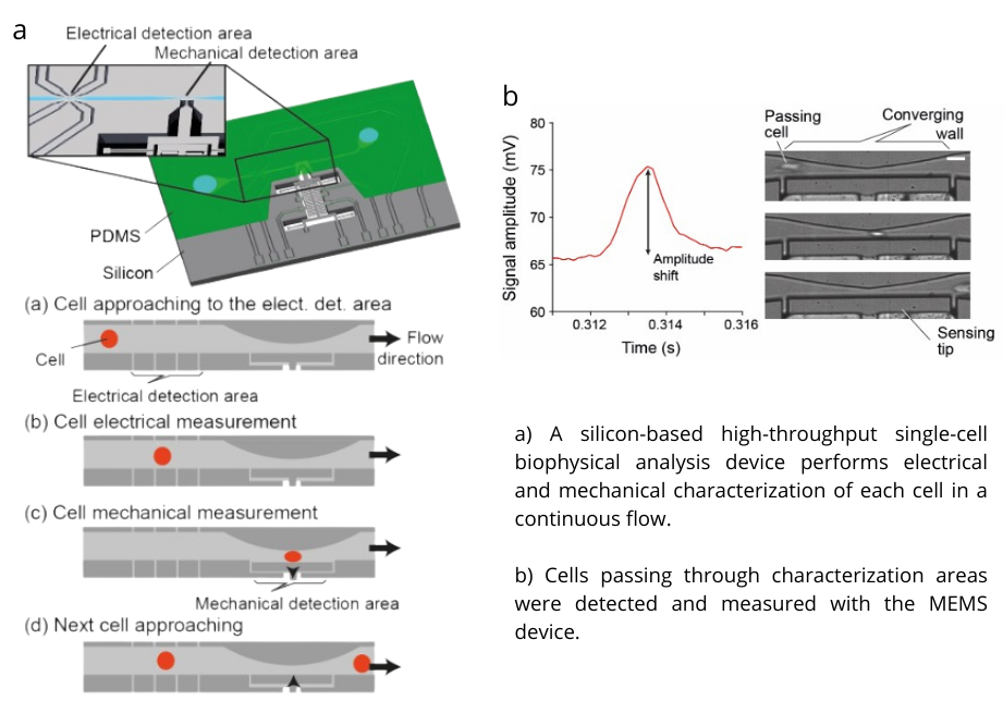
The first one is studying the biophysical properties of cells, as they have profound significance in the development of diagnostic tools and therapeutic interventions for cancer. CTCs, in particular, offer valuable insights into disease progression and treatment efficacy. We have been engaged in developing a high-throughput method for integration into routine medical assessments, and a sensitive method for versatile analysis answering fundamental questions. These devices explore the biophysical attributes of cells to discern their metastatic potential.
![]() Contact : cagatay.tarhan
Contact : cagatay.tarhan ![]() yncrea.fr
yncrea.fr
Publications ________________
(i)
- Y. Tauran, M. Kumemura, M. C. Tarhan, G. Perret, F. Perret, L. Jalabert, D. Collard, H. Fujita and A.W. Coleman, “Direct measurement of the mechanical properties of a chromatin analog and the epigenetic effects of para-sulphonato-calix[4]arene“, Scientific Reports, 9, 5816, 2019.
- Y. Takayama, G. Perret, M. Kumemura, M. Ataka, S. Meignan, S. L. Karsten, H. Fujita, D. Collard, C. Lagadec, M. C. Tarhan, Developing a MEMS Device with Built-in Microfluidics for Biophysical Single Cell Characterization. Micromachines, 9, 275, 2018.
- Y. Tauran,* M. C. Tarhan,* L. Mollet,* J.B. Gerves, M. Kumemura, L. Jalabert, N. Lafitte, I. Byun, B.J. Kim, H. Fujita, D. Collard, F. Perret, K. Suwinska, C. Goutaudier and A.W. Coleman, “Elucidating the mechanism of the considerable mechanical stiffening of DNA induced by the couple Zn2+/Calix[4]arene-1,3-Odiphosphorous acid“, Scientific Reports, 8, 1226, 2018, (*: equal contribution).
(ii)
On going projects ___________
(i)
- High-throughput identification of circulating cancer cells using biophysical signature, I-Site ULNE, 2018-2022.
- Modeling and classification of cancer cells using biophysical signatures, Region HdF / I-Site (PhD student support), 2020-2023.
- Smart MEMS Instrumentation for Biophysical flow Cytometry with Statistical Learning, ANR PRCE, 2022-2025.
(ii)
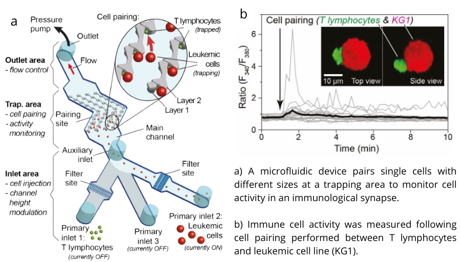
- Towards precision immunotherapies: Microfluidic single cell technology to unravel the PD-1/PDL1 calcium signature in the immunological synapse, Fondation ARC, 2019-2022.
- Role of calcium signaling in acute myeloid leukemia and the immunological synapse, PLBIO, INCa, 2023-2027.

Collaborations:

Development and understanding of innovative pharmacological approaches
High throughput opto-fluidic devices for cells treatment and therapies
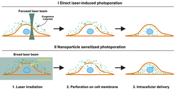 In diagnosis and therapy protocols, improving the efficiency of biological cell manipulations, like cell labelling or cancer immunotherapy, requires a controlled release of large molecular agents at high throughput. Several drawbacks characterize up to date techniques: cytotoxicity, low efficiency and throughput, specificity in drug or size or type of molecules. This project consists of developing a new high cell yield approach using gold nanoparticles mediated photoporation in a microfluidic chip, offering high throughput and low cytotoxicity drug delivery for adherent or circulating living cell. Instead of fixed plasmonic patches, we propose a new configuration with the use of advected AuNPs to allow flow photoporation. With a fully versatile setup, the spatial organization within both the biological cell and AuNP flows becomes a key point to avoid cytotoxicity due to cells/AuNP contacts.
Devices and results _________
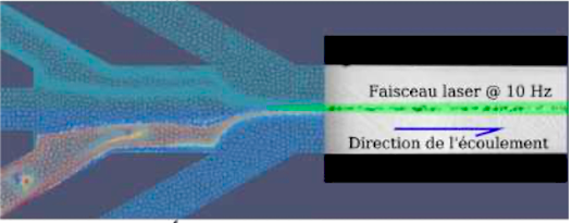 We have developed an original optofluidic devices based on a specific microfluidic design to finely control the relative distance between the flow containing the cells and the flow with gold nanoparticles. Additionally, to this improvement, tuning the distance between the two species gives us control over the mechanical stress caused by nanobubble collapse, and thus makes the optofluidic device compatible with stress sensitive cells.
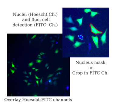 We worked through various protocols to establish a method to perform distant photoporation, that is, separating AuNPs from cells without pre-incubation in the prospect of retrieving AuNPs free photoporated samples. HeLa WT cells and AuNP suspension are injected in a microfluidic chip through separate inlets in such a way to control the distance between both distributions. Optical design was made to shape the laser beam properly and submit cells to a single pulse during their travel through the microchannel with respect to the flow rate. Permeabilization of cell membrane was monitored using FITC-dextran macromolecule incorporation via fluorescence microscopy.
Contact : anthony.treizebre ![]() univ-lille.fr
univ-lille.fr
Contact : emmanuel.courtade ![]() univ-lille.fr
univ-lille.fr
Publications ________________
- Treizebre, Q. Thommen, J. Pesez, H. Damart, L. Liegeois, E. Courtade, “Device and method for discriminating spermatozoa”, Genes Diffusion / Université de Lille / CNRS. WO2018134281 (2018/07/26), INPI : FR3061910, 2018/07/20 (BOPI 2018-29).
- A.Treizebre, Q. Thommen, J. Pesez, H. Damart, L. Liegeois, E. Courtade, « Device and Method for selecting eukaryotic cells in a transportation channel by altering the eukariotic cells by means of electromagnetic radiation », Genes Diffusion / Université de Lille / CNRS , WO2016024061 (2016/02/18), INPI : FR3024738, 2016/02/12 (BOPI 2016-06).
- Braeckmans, A. Treizebre, R.Xiong, F.Anquez, Q.Thommen, E.Courtade, M.Layachi, « Procédé et dispositif pour fournir un écoulement fluidique », Université de Gant, Université de Lille, Numéro de demande W02020221883, PCT/EP2020062084, Date de publication 2020/11/05.
On going project _________
ERC Nanobubble (Headed Pr. K. Breackmans, Univ Ghent)
![]()
Collaborations

Innovative molecular approaches to analysing and manipulating living organisms
Innovative molecular approaches to analysing and manipulating living organisms
La biologie synthétique est un domaine en plein essor qui vise à concevoir et construire des systèmes biologiques nouveaux ou modifiés pour des applications allant de la médecine à l’agriculture. Un sous-domaine important de cette discipline est celui des cellules artificielles, qui cherchent à imiter les fonctions des cellules naturelles de manière contrôlée et prévisible.
Notre thématique de recherche explore la création de cellules artificielles capables de sentir et de réagir à leur environnement grâce à des programmes moléculaires. Ces systèmes bio-inspirés sont des vésicules bilamellaires, fabriquées à l’aide de dispositifs microfluidiques dédiés que nous développons au laboratoire. Elles se composent de trois éléments principaux : un capteur (membrane modifiée répondant aux stimuli), un ordinateur (programme moléculaire à l’intérieur de la membrane) et un actionneur (diverses réponses biochimiques).
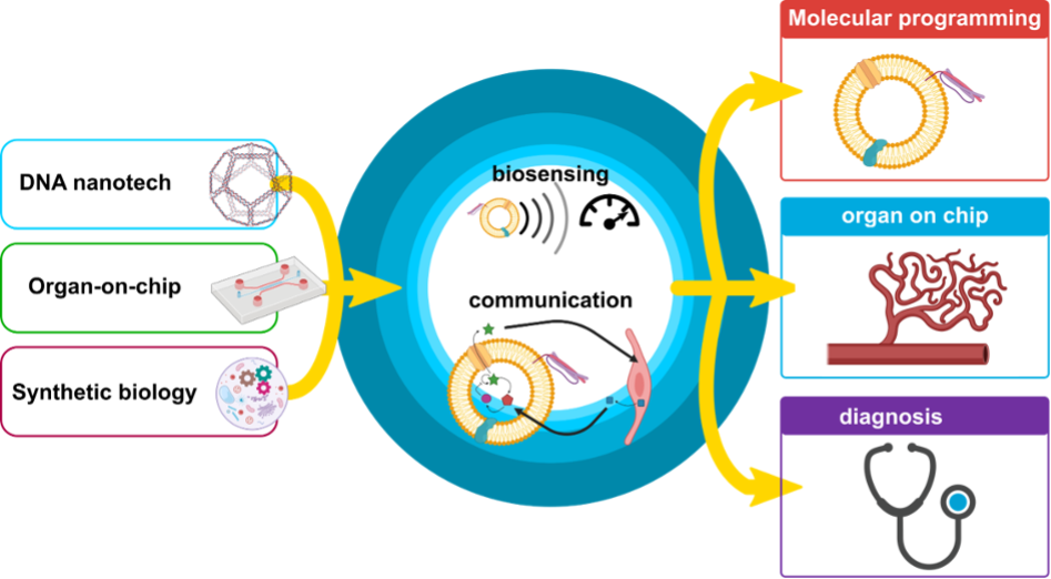
La première phase de ce projet consiste à concevoir et construire de nouveaux dispositifs microfluidiques spécifiquement pour générer ces compartiments uniques. Ensuite, les robots seront testés sur des tâches simples, comme la libération de molécules d’intérêt déclenchée par des signaux complexes ou la détection d’analytes. La polyvalence et la capacité d’adaptation de ces cellules artificielles les rendent adaptées à une large gamme d’applications, y compris des diagnostics in vitro faciles à utiliser et très sensibles.
Contact : alexandre.baccouche ![]() univ-lille.fr
univ-lille.fr
Using AI for health
Advancing Histopathological Image Analysis: AI-Driven Computer Vision for Diagnostic Classification and Treatment Response Prediction
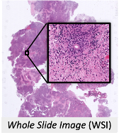 Cancer represents a major public health challenge, requiring innovations to improve diagnosis and predict responses to treatment. Our projects use artificial intelligence to analyse data such as microscopic images and thus contribute to this fight.
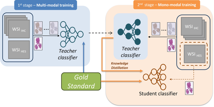 The main objective of this area of the team’s activity is to develop and refine artificial intelligence (AI) models capable of diagnosing various types and subtypes of cancer based on histopathological data. We also aim to predict patients’ life expectancy and their response to specific treatments.
Contact : feryal.windal ![]() junia.com
junia.com
Publications ________________
3- Michael Selasi DZAMESI et al. « A convolution-assisted vision transformer for the classification of Pancreatic Ductal Adenocarcinoma » CCCIE 2024, Springer.
On going projects ___________
ANR IA_Ingineering (2021-2024) https://anr.fr/Project-ANR-20-THIA-0016
Protocole Région Ethics (2023-2024). Aucun lien existant
JUNIA (2020-2021) Fond interne www.junia.com/fr/
Eurasanté (2020) (https://www.eurasante.com/appel-a-projet/innovation-prevention/)
Collaborations
Axe 2 : Conception et caractérisation de matériaux innovants :
Bio-based materials and microtechnologies for the bio-economy
Biosourced Materials and Micro-Technology for the Bioeconomy
Context:
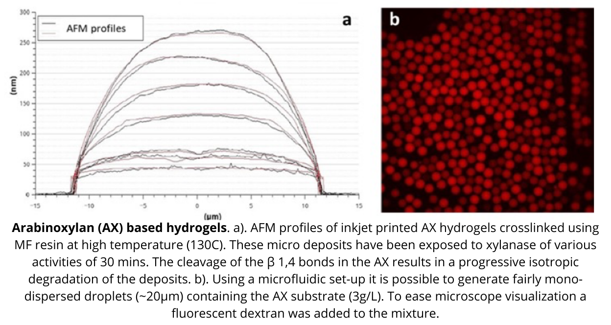
The increase in fossil resource prices, the fact that they will not be indefinitely available, and the need to act for environmental preservation drive the search for alternative technologies to valorize biomass. Thus, our team is working on developing enzyme screening tools and sensors for biomass valorization. To do so we rely on advanced microfluidics devices produced in our cleanroom, optical or electrical sensors and novel molecular approaches.
Following a series of ANR projects and technological maturation funding from SATT Nord, a startup was created in 2019: Zymoptiq, which developed an innovative sensor and method enabling simpler, faster, and more reliable enzyme analysis.
This thematic is developed through a closed collaboration with Y. Coffinier (NCM group, IEMN).
Contact : celine.vivien ![]() univ-lille.fr
univ-lille.fr
Contact : alexis.vlandas ![]() univ-lille.fr
univ-lille.fr

Non wetting Slippery infused surfaces
Non wetting Slippery infused surfaces
Context:
Developing surfaces that do not adhere to liquids has applications in a large number of day-to-day applications (vehicle windscreens, surfaces that do not adhere to molecules or viruses, anti-icing, etc.). These surfaces are generally called superhydrophobic and are obtained by multi-scale texturing (at micro and nano scales) but are sometimes not very resistant to impacts, vibrations, temperature variations or a wide range of liquids (such as alcohols). To overcome these limitations, we are working on textured surfaces that are impregnated with an inherent liquid (typically oil). The liquid then sees only a smooth surface of oil on which it can slide.

Objective: Obtain stable oil infused non wetting surfaces inside textured surfaces from Nepenthes plant bioinspiration.
Applications: Antibiofouling, anti icing, non wetting surfaces.
Challenge: Good oil retention properties by playing with both surface texturation and chemistry. Adjusted in function of application field.
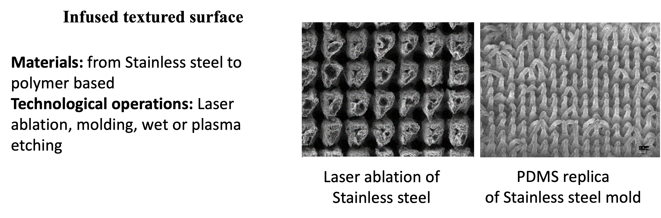
Need to realize large surface (up to 100 cm²) to authorize characterization out of the laboratory. Ex. Windscreen (transparency required), production line…
This thematic is developed through a closed collaboration with Y. Coffinier (NBI group, IEMN).
Contact : vincent.thomy ![]() univ-lille.fr
univ-lille.fr
Publications ________________
- Antifouling biomimetic liquid-infused stainless steel: application to dairy industrial processing
ZOUAGHI S., SIX T., BELLAYER S., MORADI S., HATZIKIRIAKOS S.G., DARGENT T., THOMY V., COFFINIER Y., ANDRE C., DELAPLACE G., JIMENEZ M., ACS Appl. Mater. Interfaces 9, 31 (2017) 26565-26573 doi: 10.1021/acsami.7b06709 - Manon Saget, Caroline Françolle Almeida, Vanessa Fierro, Alain Celzard, Guillaume Delaplace, et al.. A critical review on surface modifications mitigating dairy fouling. Comprehensive Reviews in Food Science and Food Safety, 2021, 20 (5), pp.4324 – 4366.
- Anne-Sophie Vaillard, Manon Saget, Flavie Braud, Marc Lippert, Laurent Keirsbulck, et al.. Highly stable fluorine-free slippery liquid infused surfaces. Surfaces and Interfaces, 2023, 42, Part A, pp.103296.
On going projects ___________
CNRS INSIS Polytic (2020)
StartAIRR Saathaf (2021-2022)
Collaborations
Smart textile for personal thermal management
Smart textile for personal thermal management
Our research theme, in collaboration with the Physics team (Ephony), aims to bring additional thermal comfort to the wearer, by adding polymer membranes to traditional textiles, thereby influencing the way in which the garments retain the heat emitted by the body. The aim of these membranes is to modify the interaction between textile and IR emitted by human body , thus accentuating the sensation of « warmth » provided by the garment, or, providing a sensation of coolness compared with an untreated textile. In the long term, a dynamic changeover between the « warm » and « cool » states could be envisaged, depending on external conditions.
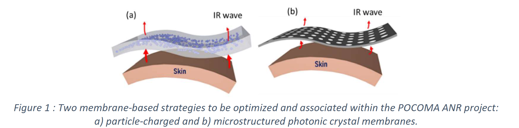
Our work over the period has focused mainly on the development of micro-structured polymer membranes to improve the thermal comfort experienced by a person at rest, by playing on the interaction between the textile and the radiation emitted by the human body in the mid-infrared (MIR) range (5-15µm).
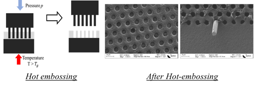
2 strategies are currently studied to achieve this microstructuring (which could also be combined): either the integration of submicrometer particles within a polymer, or its structuring as a photonic crystal on the medium-IR scale.
This technological development, initiated through the Interreg France-Wallonie-Vlaanderen PHOTONITEX project and continued by the ANR PRCE POCOMA, has not only enabled the development of a thermal model for these membranes, but also the production of laboratory-scale demonstrators which have enabled room temperatures to be reduced by 1 to 2°C, while maintaining the individual’s thermal comfort.
At present, the main challenge of the POCOMA project is to create demonstrators compatible with the textile industry by developing appropriate techniques.
Contact : Michele.carette ![]() univ-lille.fr
univ-lille.fr
Publications ________________
- , , , , , , , , , Asymmetric Design for a High-Performance Indoor Radiative Heating Fabric. Adv. Mater. Technol.2022, 7, 2101738. https://doi.org/10.1002/admt.202101738
- Polymer photonic crystal membrane for thermo-regulating textile, Assaf S., Boutghatin M., Pennec Y., Thomy V., Korovin, A., Treizebre A., Carette M., Akjouj A., Gidik, Djafari-Rouhani B., Scientific Report (2020, to be published)
On going projects ___________
This work was partially supported by :
- Damart industrial contract (2021)
- Interreg Phtonitex (2028-2022) : http://www.photonitex.eu/
- ANR PRCE (IEMN): Pocoma (2022-2026) : IEMN, DSB, GEMTEX, Junia, FOTON : https://www.iemn.fr/projet-anr-pocoma
- Phd Funding Région Hauts-de-France – Univ. Lille, collaboration Univ Mons (Be)