
1. Microfluidics for biology

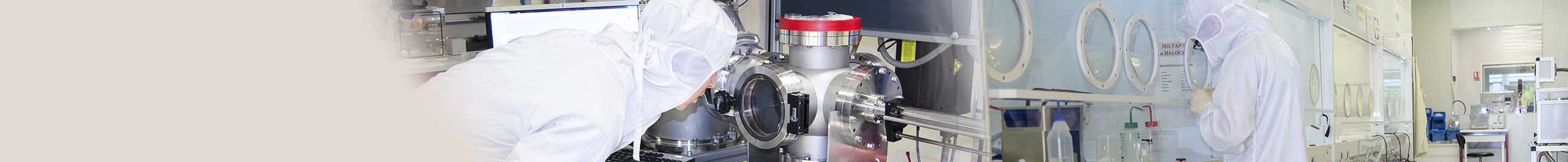
Our L2 BioMicrofluidics laboratory is a biological laboratory dedicated to the confined experimentation of biological agents classified in group 2 (French regulation of 16/07/2007) and genetically modified organisms (GMOs) to be confined in a level 2 laboratory (HCB Manual, 30/11/2014). This offers all the equipment required for cell biology (type II PSM, control microscope, automatic cell counter, CO2 chambers, etc.). This area also includes work surfaces equipped with fluid actuation systems: syringe pump (Nemesys, Harvard), pressure controller (positive/negative Push-Pull fluigent), control computers, flow meters, etc. for the flow study of micro-devices. A CO2 incubator is available in each of these work areas for microsystem cell culture.

The cluster has acquired a 3D bioprinter. Using the 3D bioprinting technique, cells and biomaterials can be deposited layer by layer to create cellular structures with properties similar to natural tissues.
2. Microscopy for biology


This area also features a microscopy room with 3 microscopes.
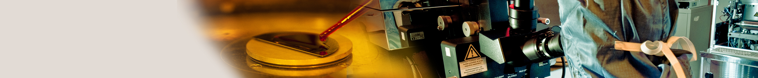
The first microscope is a Leica DMi8 with a motorised X, Y and Z stage. It can be used for long time-lapses thanks to its adaptive focus maintenance system and its environmental chamber (temperature and CO2 concentration control). This microscope is particularly well suited to real-time monitoring of cells in fluidic microsystems. A photopatterning system (Primo d'Alvéole) is also installed on the Leica DMi8. This can be used to micropattern proteins and generate any pattern with several proteins on conventional cell culture media in just a few seconds.
A second Echo Revolution microscope from the BiCo Company is a patented microscope that can be switched from upright to inverted configuration in just a few seconds by simply tilting the microscope body. This is particularly useful for inverted microscopy (for live cell samples in multi-well format, flasks, petri dishes, etc.) but also for upright microscopy (for fixed samples in particular). It is equipped with an environmental chamber for real-time monitoring of live cell experiments. It can also be used for time-lapse, stitching, multi-channel and 3D experiments.
A third microscope is a BC43 confocal benchtop from Andor. This can be used for fluorescence microscopy, transmitted light, confocal imaging and super-resolution. The Imaris introductory software installed on the microscope workstation can be used to view image stacks in 2D/3D and 4D images. This software can be used to create high-resolution snapshots and multi-dimensional films. This microscope is also equipped with an environmental chamber.
3. Molecular biology

The molecular biology platform is equipped with tools for the quantification of nucleic acids/proteins
Implen™ NanoPhotometer™ NP80
The Implen™ NanoPhotometer™ NP80 is a compact and intuitive spectrophotometer, ideal for the quantification of DNA, RNA and proteins. It offers accurate measurement over a wide measurement range, not to mention its ability to measure volumes as low as 0.3 µL.
CFX Opus Real-Time PCR System
The system CFX Opus Real-Time PCR is a high-performance quantitative PCR (qPCR) platform designed for research and diagnostic applications. This real-time thermocycler is equipped with optics that provide fast, accurate, multiplexed detection of 5 fluorescent signals, ensuring reliable quantification of nucleic acids. CFX Maestro software enables full data analysis, making it easy to interpret results.
Electrophoresis system
The laboratory also has aelectrophoresis for the separation and analysis of nucleic acids and proteins. The system is composed of several units, including horizontal electrophoresis tanks for the analysis of DNA fragments by agarose gel electrophoresis and vertical tanks for the separation of proteins by polyacrylamide gel electrophoresis (SDS-PAGE). The equipment is complemented by UV transilluminators and digital imaging documentation systems, enabling the display and quantification of labelled DNA or protein bands.


