New advance in the fight against one of the most aggressive paediatric cancers
Diffuse midline gliomas (DMG) are among the most aggressive and devastating paediatric brain tumours, with very rapid progression and a mortality rate of approximately 98% within one year of diagnosis. Located in deep and vital regions of the brain, they are almost impossible to remove surgically, and radiotherapy, which offers only temporary relief, remains the only standard treatment more than fifty years after its introduction.
Over the past decade, new discoveries about the genetic [1] and epigenetic [2] factors of GMD have raised hopes for new therapies. Scientists have identified specific mutations in histone genes [3] that profoundly alter the way tumour cells regulate their DNA, suggesting that “epi-drugs”, which target these epigenetic changes, may be promising. In laboratory studies, several of these drugs have shown strong anti-tumour effects. However, in clinical trials, they have failed to improve patient survival.
Why is there such a discrepancy? One of the main reasons lies in how we test therapies before they reach patients. Most preclinical models [4] rely on cancer cells grown in 2D on plastic surfaces, under conditions that are very different from the complex environment of a real tumour. Inside the brain, tumour cells live in a dense, three-dimensional matrix of proteins, surrounded by gradients of oxygen [5] and nutrients. These physical and chemical constraints strongly influence their growth, stress resistance and response to treatment.
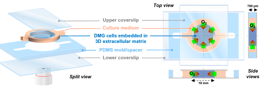
Schematic representation of the DMG-on-Chip
To fill this gap, as part of a highly interdisciplinary project led by Dr Furlan and Dr Meignan from the OncoLille Institute and the Oscar-Lambret Centre in collaboration with the IEMN and funded by the INCA, a new ‘DMG-on-Chip’ model » – a miniature, transparent device that recreates the essential characteristics of the tumour’s natural habitat.
Caption:
[1] Most cancers are caused by changes in genes that usually occur randomly and are not necessarily hereditary. They are acquired at some point in a person’s life. These genetic changes are called sporadic (spontaneous) or acquired mutations.
[2] Epigenetics is the study of the biochemical mechanisms that regulate gene expression without altering the DNA sequence. In fact, it is not always the sequence that matters, but how it is used. The link between epigenetics and cancer stems from epigenetic dysregulation (histone acetylation, DNA methylation, etc.) which, for example, can inactivate tumour suppressor genes that are naturally present in the genome.
[3] DNA-binding proteins in the cell nucleus. Each strand of DNA, approximately 50 nanometres long, is typically wound into small ‘beads’ around a block of eight histones, resulting in a ‘string of beads’ structure called chromatin, which forms the basis of the structural organisation of chromosomes. This dense organisation allows the entire length of human DNA, approximately 1.5 metres when uncoiled, to be compacted into the cell nucleus, a spheroid with a diameter of approximately 10 micrometres.
[4] A cancer model is an artificially induced or natural system that shares characteristics with human malignant tumours, for example isolated cells or laboratory animals. Preclinical studies aim to determine the potential efficacy of a drug, procedure or treatment and must be carried out before any human trials (regulated in France by Law 2012-300 of 5 March 2012 on research involving human subjects, and subsequent amendments).
To address this gap, a new ‘DMG-on-Chip’ model – a miniature, transparent device that recreates the essential characteristics of the tumour’s natural habitat – has been developed. In this microfluidic chip, [6] a 3D tumour is embedded in a gel containing key components of the brain’s extracellular matrix, such as collagen, laminin and hyaluronic acid. Oxygen is supplied only at the edges, creating a gradient similar to what occurs inside real tumours: well-oxygenated cells at the periphery and stressed, oxygen-deprived cells at the centre.
Unlike conventional ‘passive’ cell cultures, this configuration allows scientists to monitor in real time how tumour cells behave under these realistic conditions, and to continuously modify and adjust the experimental conditions. As oxygen levels drop, brain cells slow down their division, rewire their metabolism and activate genetic programmes that help them survive. Using advanced imaging and transcriptomic analyses [7], the researchers mapped these changes on the chip and discovered the molecular signatures of stress and resistance that occur when brain cells are hypoxic.
The platform also allows for more precise testing of the effects of therapies. By analysing how different regions of the chip respond to drugs or radiation, we were able to reveal striking spatial heterogeneity: while some cells die, others – protected by their microenvironment – survive and continue to grow. Such trends seem to reflect what clinicians have observed in patients and may help explain why some treatments fail, even when they appear effective in standard laboratory tests.
The immediate application to DMG, the DMG-on-Chip approach developed with the IEMN, represents a transformative shift in cancer research: moving towards more realistic and interdisciplinary models that integrate biology, physics and engineering, as well as mathematical modelling.
By faithfully reproducing the biophysical conditions of a tumour, this model could accelerate the arrival of more effective treatments for these children, who currently have virtually no therapeutic options. More than just a scientific tool, DMG-on-Chip could well become an indispensable link between the laboratory and the patient’s bedside, helping to transform one of the most tragic childhood diseases into a field of hope and progress.
Caption:
[5] The word ‘gradient’ is the mathematical definition of the spatial variation of a measured quantity, in this case the variation (decrease) in oxygen concentration in tissues. Hypoxic brain injury is damage to certain parts of the brain resulting from insufficient oxygen supply to the affected area. The decrease in oxygen supply to tissues, called hypoxia, damages cells and can lead to their destruction.
[6] The term microfluidics encompasses all the sciences and techniques of systems that manipulate fluids in circuits manufactured by printing on transparent materials (allowing access and visualisation), at least one of whose characteristic dimensions is in the order of a micrometre. These devices can handle microscopic quantities of fluid (billionths of a litre) comparable to the size of cells.
[7] Transcriptomics is a sub-discipline of genomics devoted to the study of the transcriptome, i.e. all the RNA molecules (transcripts) resulting from the transcription of DNA comprising the entire genome of an organism. Transcriptomics emerged with the advent of high-throughput analysis techniques such as DNA microarrays and, more recently, high-capacity DNA sequencers. Comparing transcriptomes makes it possible to identify genes that are differentially expressed between distinct cell populations or in response to different treatments.


