Nanopuces à ADN pour appréhender les cellules tumorales
Detecting metastatic cancer cells circulating in blood vessels is a very difficult task. It involves identifying a serial killer hidden among billions of individuals, diluted in a few drops of blood. The criminal's fingerprints are certain proteins found on the surface of the circulating tumour cell. Fortunately, small DNA sequences called aptamers are capable of uniquely recognising them, even in minute quantities. We have designed nano-chips that align thousands of these 'police' aptamers, which can stick to the tumour proteins in a liquid biopsy and identify the killer cell among the millions of normal cells, with a characteristic electrical signal.
One of the main causes of death in cancer patients is organ failure induced by the development of metastases. The primary tumour spreads throughout the body by generating circulating tumour cells (CTCs), i.e. cancer cells that detach from the tumour and circulate in blood vessels. Our institute, the IEMN, is a partner in an international, multidisciplinary research effort aimed at developing an original and innovative family of lab-on-a-chip devices designed to detect CTCs with high efficiency. The devices are based on a sophisticated quantum mechanical effect that enables the presence of a cancer cell immobilised on the chip to be detected with extreme precision. Briefly, a DNA fragment carrying a redox molecule at one end is sandwiched between two electrodes; at equilibrium, the DNA fluctuates randomly and the redox molecule induces a very weak current, via the quantum tunneling effect of electrons from/to the negative electrode; in the presence of a CTC, the DNA is able to identify the presence of cancer-specific antigens present on the surface of the CTCs, whose presence is detected by the variation in the quantum tunnel current. The first results of this technique were published recently in the journals Biosensors and Bioelectronics and ACS Sensors. In what follows, we give a fuller description of the technique and its applications. main results.
The first protagonist of this story is the epithelial cell adhesion molecule (EpCAM). This protein, present on the outer membrane of epithelial cells, has received much attention as the main membrane marker used to isolate CTCs. The biology of EpCAM and its role are not fully understood, but evidence suggests that expression of this epithelial cell surface protein is crucial for metastatic CTCs, given that most cancers arise from epithelial cells. A typical outcome of clinical screening is that progression-free survival and overall cancer survival times are significantly reduced in patients with ≥5 CTCs per 7.5 ml blood sample, or ≥20 EpCAM-positive EVs. Consider that such a volume of blood contains around 40 billion red blood cells and around 50 million other cells (leukocytes, granulocytes), and you can get an idea of the level of resolution required to capture and discriminate a single CTC from the entire normal cell mass.
The other main characters in the play are the DNA aptamers. Aptamers are short fragments of artificially synthesised DNA or RNA (ssDNA or ssRNA) that can bind to a specific target protein, as well as peptides, carbohydrates, small molecules, toxins and even living cells, with extreme selectivity in the nM range (meaning they can selectively find a target element in a sample of a billion). Since their discovery in the early 1990s, considerable efforts have been made to make them clinically relevant for diseases such as cancer, HIV and macular degeneration. With the advances in high-precision medicine, targeted therapy, imaging and nanotechnology, aptamers are readily being considered as the best potential targeting ligands, thanks to their inexpensive chemical synthesis and the ease with which they can be modified to suit variable targets.
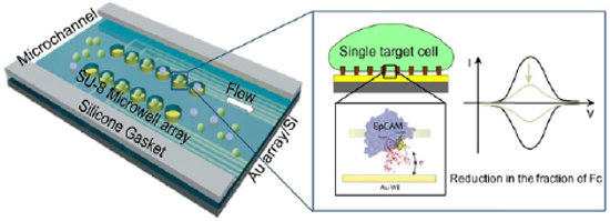
If the DNA aptamers and EpCAM found on the surface of the CTCs are two lovers to be wed, here's the Reverend Father conducting the ceremony. This is a variant of the electrochemical method of cyclic voltammetry (CV), a fairly old and standard experimental technique that almost anyone can run in their garage. In its most basic configuration, CV detects the presence of molecules capable of transferring electrons, i.e. species of redox which can be reduced or oxidised; by periodically varying the potential of the negative electrode, so as to scan the energy levels of the redox molecule, electrons can be transferred to the molecule and vice versa, generating a current. The shape of the plot of potential versus current is very typical of each molecular species and describes the energy barrier that electrons must cross to move from one stable state to another. Here we transform the general CV method into an extremely sophisticated microscopic diagnostic tool. In our experiments, a redox molecule is attached to one end of a short DNA aptamer, which in turn is attached to a gold electrode at the other end. In this configuration, the DNA acts as a tether, causing the position of the redox molecule above the electrode surface to fluctuate randomly between a minimum approach distance and a maximum elongation distance. When the molecule fluctuates close to the surface, the electron can pass through the barrier by quantum tunneling and give rise to a small but measurable current; but when the molecule fluctuates far from the surface, the current becomes zero. This is the key operating principle of our innovative CTC detection method: because the DNA aptamer sequence is designed to selectively recognise the EpCAM protein, when a CTC cell is present 'above' the electrode in solution, the DNA aptamer sticks to it, blocking the redox species at a position well above the electrode surface, and effectively cutting off the current signal.
The current device is a microfluidic lab-on-a-chip, just a few millimetres in size.2 (see figure). The surface of the Au electrode is decorated with a regular array of plastic nanopillars (cross-linked hydrogen silsesquiloxane), 20 nm high and 200 nm wide, spaced 500 nm apart. The DNA aptamer attached to the Au surface has an extended length of about 15 nm, but at room temperature it adopts a partially folded conformation, reducing it to a more compact structure of about 10 nm; a ferrocene molecule (redox species) is attached to the free end of the DNA. The ferrocene-bearing DNA aptamers are deposited as a dense monolayer on the Au surface. The whole device is integrated into a microfluidic chip, in which a solution containing different types of target cells is transported at low speed. In initial experiments, CAPAN-2 pancreatic cancer cells and RAMOS Burkitt's lymphoma cells were used as a test. The moving cells are trapped on the nanopillars and remain suspended, slowly falling towards the surface under gravity. This allows surface membrane proteins, including the target EpCAM, to approach the fluctuating DNA attachment. Once each DNA is attached to an EpCAM, this contributes to a reduction in the total current measured, in the order of microamperes. The current lower limit of resolution is around 3,000 cells per ml, which is still high compared with the target. However, this first version of the technical device has considerable room for improvement and is very promising because of its versatility (the target proteins can be easily modified by changing the sequence of the DNA aptamer, different proteins can be targeted simultaneously by using several aptamers in parallel), and also because of its very low manufacturing cost compared with current standard technologies.
The team is made up of a multidisciplinary group of physicists, chemists, engineers and biologists from the LIMMS laboratory in Tokyo (Japan), the IEMN and the CANTHER cancer research unit, both in Lille (France). The contribution of the IEMN team focuses on on electrode surface chemistry and DNA immobilisationled by Dr Yannick Coffinier, and on the computer simulation of DNA-protein interactions in solution, directed by the Prof. Fabrizio Cleri .


