
Conventional ultrasonic detection and imaging methods (such as medical ultrasound or industrial NDT) are based on the transmission and reception of ultrasonic echoes reflected once at the interfaces present in the medium. By analysing these echoes, it is possible to obtain location information or to produce images, provided that the interfaces or objects in question have acoustic properties that are significantly different from those of the surrounding environment (which explains, for example, the fact that in obstetric ultrasound, it is the baby's skeleton that presents the best contrast on the images).
In contrast, in the application presented here, we use ultrasonic multipaths (echoes reflected a very large number of times on the different interfaces of the propagation medium). Unlike ultrasound, this technique does not allow imaging (since the location information is lost), but it is extremely sensitive to even small variations in properties. These multiply reflected echoes are known as "coda signals", from the Latin word coda meaning tail (they appear at the end, or "tail" of the recorded ultrasound signals).
From these ultrasonic codas, a so-called "decorrelation" coefficient is calculated. This coefficient, with values between 0 and 2, reflects the degree of dissimilarity between two signals: 0 corresponds to identical signals and 2 corresponds to unrelated signals. In the absence of any change, coda signals are perfectly reproducible from one measurement to the next. This means that the decorrelation coefficient between one of these signals, measured at a given time, and a so-called reference signal, measured at an earlier time, is close to zero.
Conversely, when a change in properties occurs at the substrate-medium interface, variations in the reflection of ultrasound waves modify the coda signals. This results in increasing values of the decorrelation coefficient. Regular measurement of this coefficient enables the evolution of a medium or interface to be monitored over time.
We have used this principle to detect the appearance of a bacterial deposit (biofilm) in a culture medium contained in a tank. The ultrasonic sensor is glued to the outer surface of the tank and is therefore never in contact with the medium. The ultrasonic measurement is made through the wall.
The value of the correlation coefficient between the signal measured at the initial instant and the successive signals at each subsequent instant could be directly correlated to the growth of the bacterial population, as shown in Figure 1. In this figure, graph A shows the evolution of the decorrelation coefficient as a function of time when the tank contains a sterile medium (absence of bacteria and therefore no growth of biofilm on the internal walls of the tank). Graph B shows the evolution of the coefficient in the presence of bacteria in the medium. Comparison of these two graphs clearly shows that the decorrelation coefficient is very sensitive to the growth of the bacterial population, which leads to the formation of a biofilm on the wall. This is confirmed by a measurement of the bacterial concentration over time (graph C).
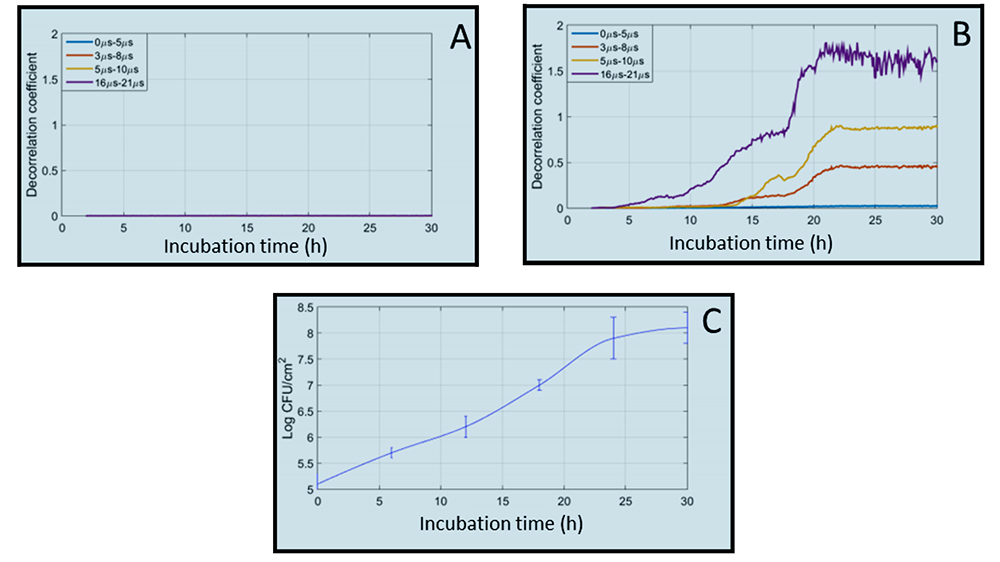
Figure 1: Change in decorrelation coefficient as a function of incubation time and comparison with bacterial growth kinetics. (A) Control sample (sterile medium). (B) sample containing bacterial culture medium (C) biofilm formation kinetics (bacterial concentration
This method seems particularly promising for non-invasive real-time on-site monitoring of biofilm formation and, more generally, of interface properties (fouling, various deposits, variations in the properties of the medium, etc.).
One of its limitations is its high sensitivity to temperature, which can lead to false positives. This is not a problem in cases where the experimental conditions are thermostatically controlled. In other cases, methods to compensate for the influence of temperature variations on coda decorrelations are currently being studied.
This method is currently the subject of the Interreg-2-Mers SOCORRO project, in which the UPHF (IEMN), INRAE and the University of Lille (UMET) are collaborating.