
Yuki TAKAYAMA
Wednesday 28 March 2019 at 2.00 pm
Amphitheatre of the IEMN-Laboratoire central - Villeneuve d'Ascq
Jury:
- D. COLLARD, IEMN / CNRS (Thesis supervisor)
- C. DEJOUS, IMS, University of Bordeaux (Rapporteur)
- V. TALY, Sorbonne Paris Cité University / INSERM, CNRS (Rapporteur)
- C. LEGALLAIS, Université de Technologie de Compiègne / CNRS (Examiner)
- C. LAGADEC, CPAC, University of Lille / INSERM (Examiner)
- M.C. TARHAN, ISEN Lille / YNCREA (Examiner)
- H. FUJITA, IIS, University of Tokyo (Invited)
Summary:
As tumour masses develop, cancer cells acquire a specific phenotype that makes them resistant to treatment and enables them to invade surrounding tissues, the peripheral bloodstream and spread throughout the body. Surgical treatment of primary tumours is mostly effective, but targeting tumour cells spread to distant organs remains extremely complex. As a result, ~90 % of cancer-related mortality is due to tumour development at secondary sites: metastases. The change in the phenotype of the cell required for metastatic progression is also accompanied by a significant reduction in its rigidity, enabling it to insert itself into the surrounding tissues. As a result, the characterisation of biomechanical properties may prove to be an original approach for detecting cancer cells in the bloodstream (circulating tumour cells).
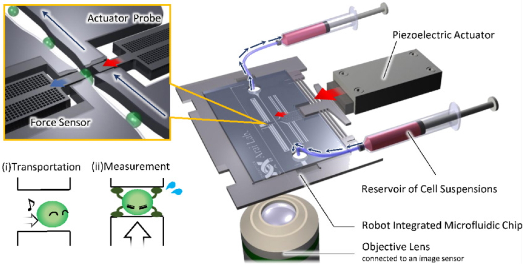
Overview of the on-chip measurement system for cellular mechanical characteristics
Recurrently, the various techniques are exposed to a compromise between the characterisation throughput (number of cells processed in a given time) and its content (number and precision of parameters measured). Some techniques, such as atomic force microscopy (AFM), enable very precise measurements but suffer from an extremely low characterisation throughput, while methods based on microfluidics, similar to flow deformability cytometry, enable interesting measurement throughputs but suffer from a lack of content, making it impossible to precisely identify the different cell phenotypes.
Circulating cells are micron-sized (10-30 microns in diameter), so microtechnologies offer multiple dimensional advantages for manipulating, stimulating and characterising them individually. MEMS (Micro Electro Mechanical Systems) are therefore ideally suited to measuring the various biophysical parameters and mechanical and electrical properties of single cells. However, MEMS devices cannot be used in a conductive liquid environment for electrical and mechanical characterisation. To overcome this limitation, this study proposes to separate the part that manipulates the cell in its biological environment from the MEMS actuators and sensors that perform the electrical and mechanical measurements. In addition, to increase the characterisation throughput and ensure operability, the MEMS device directly integrates the microfluidic channel in which the cells will circulate.


