Ultra-sensitive detection and manipulation of nano-mechanical vibrations
To date, various technologies have been developed in both industrial and research settings to detect and manipulate tiny mechanical vibrations at the nanometre scale, facilitating sensing applications. However, there is still a lack of tools that combine high spatial resolution with the ability to manipulate the energy exchange between different vibrating elements without the need for mechanical contact. Such a tool is essential for exploring extremely small energy variations, for example for sensing in quantum systems.
In this work, we present a unique and ultra-sensitive platform that uses a scanning metallic tip capacitively coupled to an underlying membrane, with no mechanical contact. Low-power microwave signals are transmitted through the tip to an underlying membrane, serving as an information bus. These microwave photons facilitate coherent interactions between the tiny mechanical vibrations of a few nanometres carried by the tip and the membrane, but also provide an ultra-sensitive method to readout this kind of interactions, with detection floor of 2.1 pm⁄√Hz. The basic concept of this platform, along with the suspended membrane fabricated using nanofabrication technologies [1], are shown in Fig.1. This setup combines high spatial resolution with the ability to inject microwaves, using the scanning tip as a movable top-gate positioned above the suspended vibrating membrane. The ultra-sensitivity allows to detect membrane vibrations even when the distance (h) between the tip and the membrane is relatively large, such as h = 1 μm. It enables the study of nanomechanical phenomena, such as mapping mechanical vibration modes (see Fig.2.(a)) and analysing mechanical damping effects. The spatial resolution can reach the nanometre scale.
Figure (1) :
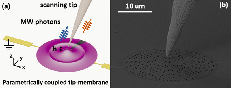
Fig.1: The graph presents a coupled scanning tip and a suspended membrane. (a) A schematic of the basic concept of the experiment shows a scanning tip positioned above a membrane microelectromechanical resonator (shown in purple), with a separation distance h along the z-axis. (b) Scanning Electron Microscopy photo of the scanning tip and the membrane. The tip has a head approximately 2 µm in size. The membrane is about 30 µm in diameter, with a thickness of around 80 nm, and is coated with a thin aluminium film roughly 20 nm thick [1]. The entire membrane is separated from the silicon substrate by etching away the silicon layer underneath through those patterned nano holes, which are approximately 200 nm in diameter [1].
Figure (2) :
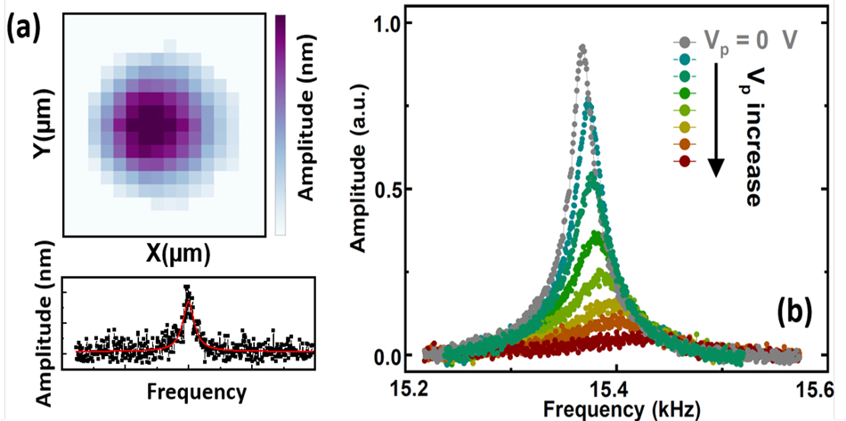
Fig.2: The graph presents unique features of this coupled tip – membrane system. (a) The spatial vibration modes of the membrane are mapped by scanning the tip across the x-y surface while maintaining a constant distance h between the tip and the membrane. The lower figure displays the typical mechanical response of the membrane when the tip is positioned at the center of the circular membrane, measured via microwave photons emitted from the tip. The upper figure presents a spatial pattern map of the membrane vibration mode, derived from the resonances measured at each x-y position. The imaging mode map is consistent with mechanical vibration theory. (b) The vibration amplitude of the scanning tip varies depending on the electrical signal Vp, which biases the interaction between the membrane and the tip.


