A nano-radar for cell imaging: 3D microwave microscopy and fluorescence in a liquid medium
The electrical activity of cellular organelles such as mitochondria plays a key role in biology and medicine, but remains poorly understood. Its link with aging, apoptosis and diseases such as cancer and diabetes raises essential questions. Developing a calibrated, broadband electronic interface would enable to explore these phenomena and pave the way for new therapeutic approaches.
Early work
In 2017, a consortium comprising the IEMN, the University of California Irvine, the Center for Mitochondrial and Epigenomic Medicine at the Children’s Hospital of Philadelphia, USA, carried out the first-ever imaging of living mitochondria using scanning microwave microscopy. Mitochondria, isolated from cultured HeLa cells, are attached to a graphene support and kept alive by a respiratory buffer that supplies them with the nutrients required for the Krebs cycle. The organelles are analyzed by capacitive measurement at a frequency of 7 GHz.
A nanoelectronic broadband interface inside living cells with integrated fluorescence readout of metabolic activity.
In 2020, the consortium achieves a major breakthrough by presenting the very first calibrated broadband electrical connection inside a living cell with integrated fluorescence readout of metabolic activity. On-chip, nanoscale capacitance calibration standards are used to quantify microwave response with cellular images obtained at 22 GHz. Such an interface opens up numerous prospects for the integration of life sciences with nanoelectronics, notably for electronic testing of membrane potential dynamics, nanoelectronic activation of cellular processes, as well as nano-radar tomographic imaging of the morphology of vital organelles within the cytoplasm, throughout the cell life cycle, in different physiological environments and under various pharmacological conditions.
Combined 3D coaxial microwave microscopy and super-resolution fluorescence: Proof-of-concept imaging of living cells in liquid media – Towards a biological nano-radar
In 2024, the same consortium is continuing its work by developing a new proof-of-concept for 3D microwave microscopy combined with super-resolution fluorescence. This new version uses micro- or nano-coaxial probes to overcome the problem of parasitic coupling. The coaxial architecture improves the spatial resolution and sensitivity of measurements by reducing unwanted absorption of the microwave signal by the biological medium. These advances lay the foundations for a true biological nano-radar capable of probing the electromagnetic dynamics of cellular organelles in liquid media, in real time.
To be continued
In January 2025, METAS, the Swiss Federal Institute of Metrology, joined the consortium, contributing its expertise in the development of nano-coaxial probes. These new probes, based on advances in nano-fabrication, are currently being integrated. Their deployment aims to extend the capabilities of the device, both in terms of spatial resolution and spectral coverage. In addition, this work is paving the way for the design of reference metrological equipment, designed to guarantee the traceability and calibration of nanoscale microwave measurements in biological environments. These developments also open up new perspectives in radio-frequency quantum detection, currently being optimized. The aim is to explore previously inaccessible measurement regimes, with enhanced precision and stability.
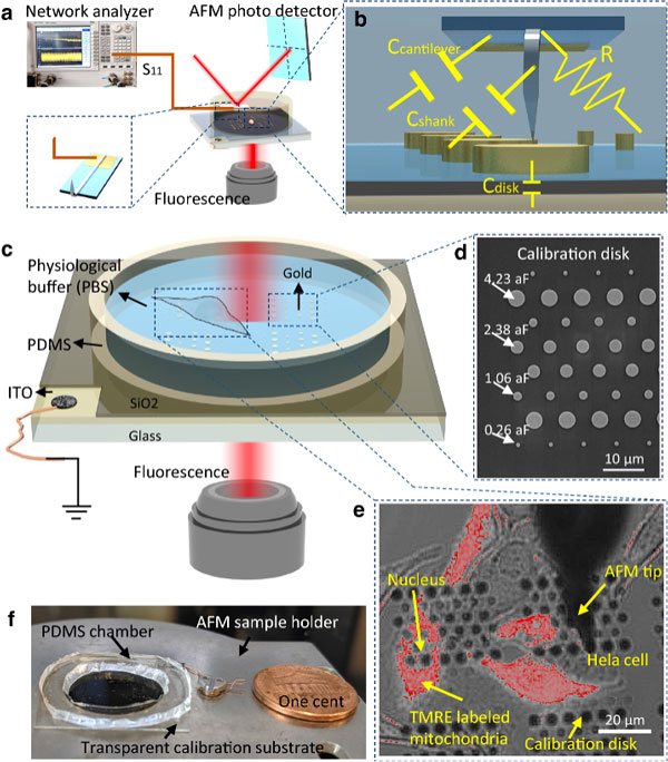
(a) A microwave vector network analyser (Keysight N5222A PNA) measures the signal reflected from a metal AFM tip. A standard AFM scanner is used to move the AFM tip over the sample under study. (b) The equivalent electrical circuit at the tip of the probe consists mainly of the capacitance between the tip and the ground plane, which varies as the tip is moved. However, unwanted parasitic elements are also present. Although these are assumed to be constant when the tip is scanned, they need to be calibrated in order to obtain a corrected image. (c) Sample chamber containing live cells and calibrated standards, together with the optically transparent electrical ground plane (ITO). (d) SEM image of the calibration discs. (e) Superimposed bright field and fluorescence image of a live HeLa cell culture. The fluorescent marker TMRE is used to indicate mitochondrial membrane potential. (f) Photograph of the sample chamber
Microwave microscopy imaging of a vital HELA cell in tapping mode
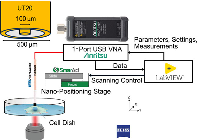
Implementation of the system for the proof of concept of coaxial 3D microwave microscopy combined with high-resolution fluorescence.
(a) Broadband measurement above a metal plate with a separation distance set at 80 μm. (b) Complex reflection coefficient S11 measured as a function of absolute Z position for the test frequencies 3.75375 GHz, 3.99335 GHz and 5.76040 GHz (MUT = HeLa cells [ATCC CCL-2] in a physiological buffer, ZSTEP = 50 μm). IFBW = 100 Hz.
References
[1] Li, Jinfeng, Zahra Nernati, Kamel Haddadi, Douglas C. Wallace, and Peter J. Burke. « Scanning microwave microscopy of vital mitochondria in respiration buffer. » In 2018 IEEE/MTT-S International Microwave Symposium-IMS, pp. 115-118. IEEE, 2018. https://hal.science/hal-03224648v1
[2] Ren, Dandan, Zahra Nemati, Chia-Hung Lee, Jinfeng Li, Kamel Haddadi, Douglas C. Wallace, and Peter J. Burke. « An ultra-high bandwidth nano-electronic interface to the interior of living cells with integrated fluorescence readout of metabolic activity. » Scientific reports 10, no. 1 (2020): 10756. https://hal.science/hal-03224644v1
[3] Lee, Chia-Hung, Kamel Haddadi, and Peter J. Burke. « Combined Super-Resolution Fluorescence and Coaxial 3-D Scanning Microwave Microscopy: Proof-of-Concept In-Liquid Live-Cell Imaging: Toward a Biological Nano-Radar. » IEEE Microwave and Wireless Technology Letters (2024). https://hal.science/hal-04815101v1
[4] Kamel Haddadi, IEEE Member, Clément Lenoir, Mohamed Sebbache, Chia-Hung Lee, Peter Burke, IEEE fellow. « Microwave Imaging with Open-Ended Coaxial Probes. » IEEE 2024 International Conference on Manipulation, Automation and Robotics at Small Scales (MARSS), Delft, Netherlands, (2024). https://hal.science/hal-04946860v1
Contacts:


